Case review · Orbit
Case review: one-piece orbitozygomatic reconstruction
Three regions, one piece: this case rebuilds the orbital floor, the lateral rim and the body of the cheekbone at once. Here is why a one-piece implant was chosen, and how it is fixed.
1 min read2 sources
The case
Defect combining the orbital floor, the lateral orbital rim and the body of the zygoma, the cheekbone. In such combined injuries, the reconstruction must both support the orbital contents and restore the contour of the cheek.
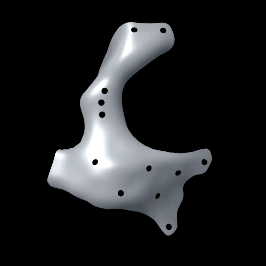 Orbit and zygomaOrbitozygomatic reconstructionFloor, lateral rim and body of the zygoma
- Overall size
- 40 × 38 × 64 mm
- Thickness
- ≈ 0.45 mm
- Volume
- 1.1 cm³
- Mass (Ti-6Al-4V)
- ≈ 5 g
The shape: mirroring the healthy side
A curved and very thin floor
The floor follows the curve rising towards the back of the orbit. Its median thickness of about 0.45 mm is enough thanks to the strength of titanium, and keeps the implant from taking up space in the orbit.
Fixation: frontal and malar
Fixation is placed on solid bone: above, on the rim towards the frontal bone, and below, on the malar bone. The lateral strut joins the two and restores the outline of the orbit.
What to look at in the 3D viewer
- Front view: comparison of both orbits, reconstructed side and healthy side.
- Coronal plane through the orbit: curvature of the rebuilt floor.
- Measurement: width of the floor, height of the lateral strut.
Sources
Documents accessed in October 2026. Amounts are given in the currency and for the year of each source.
- 1Pietzka S, Wenzel M, Winter K, et al. Comparison of anatomical preformed titanium implants and patient-specific CAD/CAM implants in the primary reconstruction of isolated orbital fractures: a retrospective study. J Pers Med. 2023;13(5):846. doi.org
- 2Kotecha S, Ferro A, Harrison P, Fan K. Orbital reconstruction: a systematic review and meta-analysis evaluating the role of patient-specific implants. Oral Maxillofac Surg. 2023;27(2):213-226. doi.org
General information: this article does not replace your surgeon’s advice. Prices vary with the patient, the hospital and the implant; only a formal quote is binding.
A cranioplasty case to review?
Surgeons: send the CT scan for a feasibility study. Patients: your section explains the care pathway.