Case review · Orbit
Case review: orbital roof reconstruction in oculoplastic surgery
The orbital roof separates the eye from the brain. Rebuilding it takes a thin implant placed under the upper rim, and fixation that threatens neither the eye nor its muscles. Here is how this case was planned.
1 min read
The case
Reconstruction of the orbital roof as part of oculoplastic surgery. The roof is the upper wall of the orbit: above it lies the anterior cranial fossa, below it the eye and its muscles.
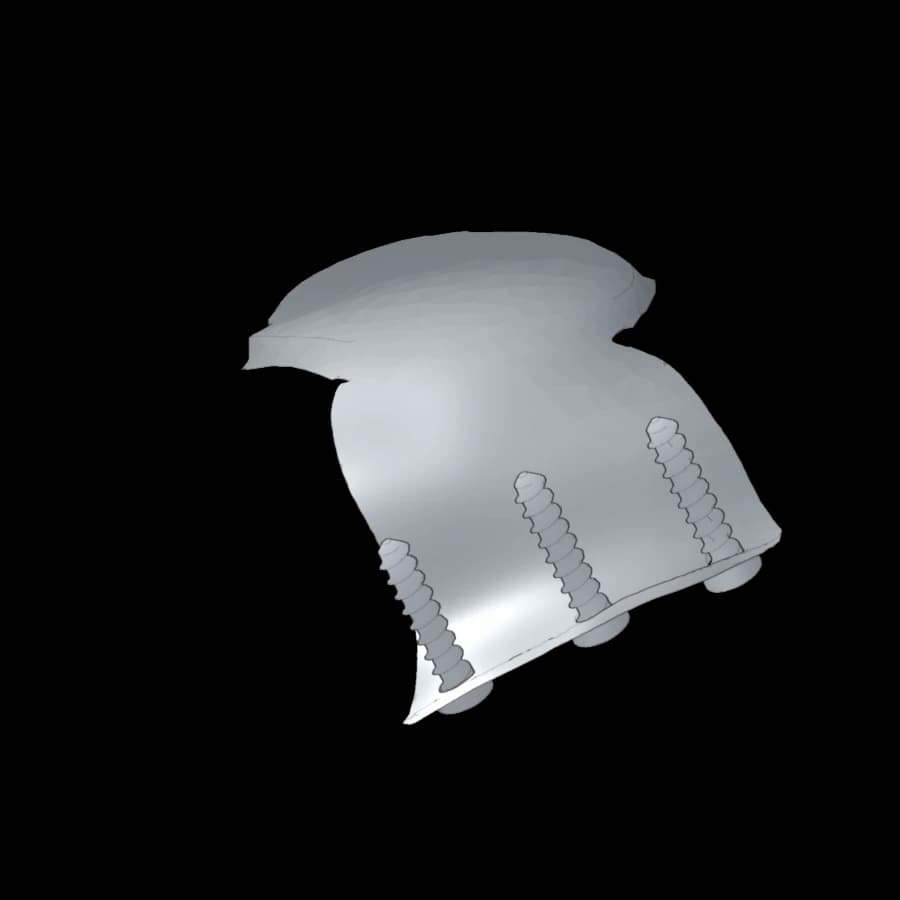 Orbital roofOrbital roof reconstructionOculoplastic surgery, fixation on the superior rim
- Overall size
- 28 × 21 × 12 mm
- Thickness
- ≈ 0.6 mm
- Volume
- 0.26 cm³
- Mass (Ti-6Al-4V)
- ≈ 1.2 g
A thin implant under the rim
The implant rebuilds the wall by following its curvature, with a median thickness of about 0.6 mm. Its shape and depth in the orbit are set on the CT scan, before surgery.
Fixation moved to the rim
Rather than screwing inside the orbit, fixation is moved to the thicker upper orbital rim. Three screws 1.5 mm in diameter and 8 mm long, 6.2 mm apart, follow trajectories set during planning, away from the orbital contents.
Why plan the trajectories
The rim is narrow and close to the eye: a trajectory chosen in theatre could leave the bone. Planning the trajectories on the 3D model ensures that each screw stays in bone, away from the structures to protect, and sets the screw length in advance.
What to look at in the 3D viewer
- View from below or from the front: position of the implant under the upper rim.
- Sagittal plane through a screw: screw trajectory in the rim.
- Measurement: spacing of the screws, depth of the implant in the orbit.
General information: this article does not replace your surgeon’s advice. Prices vary with the patient, the hospital and the implant; only a formal quote is binding.
A cranioplasty case to review?
Surgeons: send the CT scan for a feasibility study. Patients: your section explains the care pathway.